

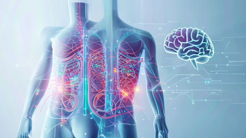
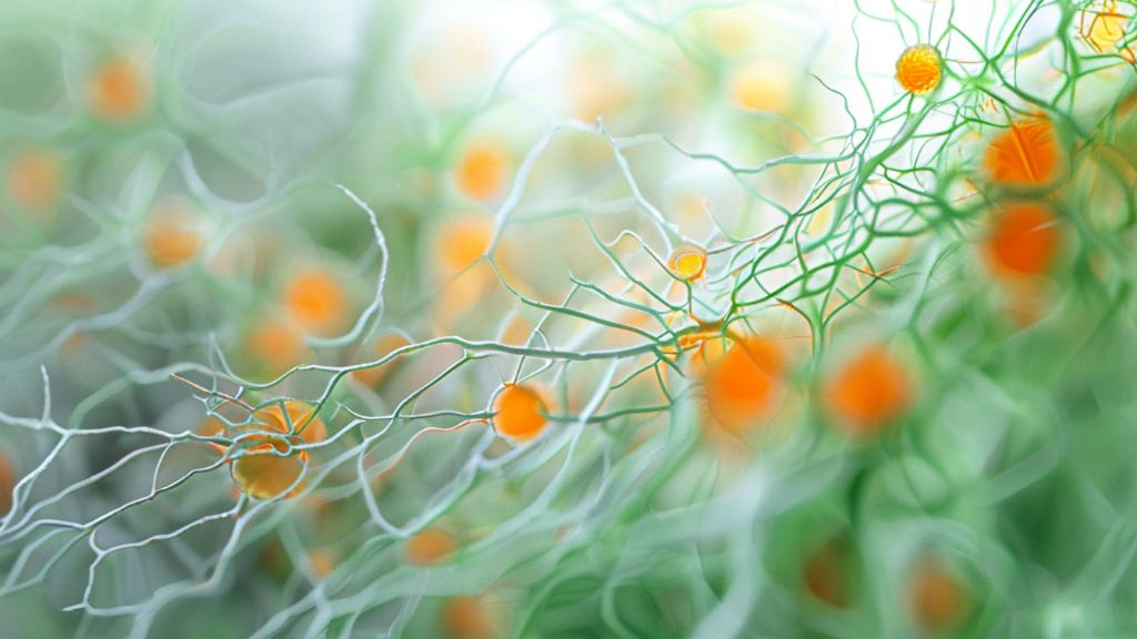
While conventional 2D pathology has been widely used for diagnosing gastrointestinal diseases, it has been challenging to accurately grasp the complex 3D structure of tissues or the dynamic living tissue environment. It's like viewing a 3D movie as a 2D photograph, making it easy to miss crucial connections or the overall picture. Sampling errors or issues arising during tissue processing can also hinder accurate diagnosis. To overcome these limitations, the need for new 3D imaging technologies has emerged. **Methodology** **Advantages** **Disadvantages** Conventional 2D Pathology Extensive research and accumulated experience Difficulty in understanding 3D structure and dynamic environment, sampling bias, potential for artifacts New 3D Technology Easy to understand tissue's 3D structure and connectivity Early stage research, requires integration of AI-based analysis 2. The Convergence of AI and 3D Imaging Technology (Introducing a New Protocol) This research proposes a method that combines 3D imaging technology with AI-based analysis to better visualize and accurately diagnose gastrointestinal tissues. It's similar to creating a complex map where you see the entire picture in 3D, and smart AI quickly identifies important information. This new protocol aims to more accurately reveal the microscopic structures of tissues and changes caused by disease. Through this, it is expected that diseases such as ulcerative colitis or Hirschsprung's disease can be better understood and diagnosed. 3. How Can We See More Accurately? (Tissue Clearing and 3D Imaging) To effectively visualize tissues in 3D, a tissue clearing process is first required. Once tissues become transparent like glass, microscopes can peer deep inside. This process involves several steps, using decolorization and electrophoretic tissue clearing methods to remove fats and other components from within the tissue. Such cleared tissues vividly show how neural structures and other important organelles are connected through 3D imaging technology. 4. How Does AI Assist Analysis? (Advantages of AI-Based Analysis) AI-based analysis processes tissues much faster and more accurately than conventional methods. For example, the analysis time for colon tissue samples can be reduced from the conventional 13 days to just 20 minutes with AI. Furthermore, AI reduces human error and increases the consistency of analysis results. Particularly, 3D measurements such as the volume or surface area of neurons can only be accurately calculated with AI. **Analysis Methodology** **Colon Tissue Sample Analysis Time** **Surgical Sample Analysis Time** **Accuracy and Consistency** Conventional Manual 2D Analysis Over 13 days 30 days Relatively low 3D Non-AI Analysis 1 day 1 day Accuracy limitations 3D AI-Based Analysis 20 minutes 4 hours High 5. How Will It Be Applied in Actual Disease Diagnosis? (Clinical Application Potential) This AI-based 3D pathology technology can be of great help in diagnosing gastrointestinal diseases such as ulcerative colitis and Hirschsprung's disease. Specifically, AI accurately distinguished areas lacking nerve cells in surgical tissues from patients with Hirschsprung's disease. In ulcerative colitis, 3D quantitative analysis of microscopic tissue changes can provide a better understanding of disease progression. Moreover, perineural invasion, an important prognostic factor in colorectal cancer, was difficult to observe with conventional 2D but can be clearly seen with AI 3D. 6. What are the Future Directions for Development? (Future of Research) These research findings demonstrate the potential of AI-based 3D pathology to bring about revolutionary changes in medical diagnosis and patient treatment. Moving forward, plans include applying this technology to a wider range of diseases and further developing AI algorithms to enhance accuracy. Combining AI analysis results with clinical data could also contribute to establishing personalized treatment strategies. Ultimately, this technology is expected to play a crucial role in diagnosing diseases faster and more accurately, and in discovering new clinical features cost-effectively. Leica AiVIA Used Paper file attached